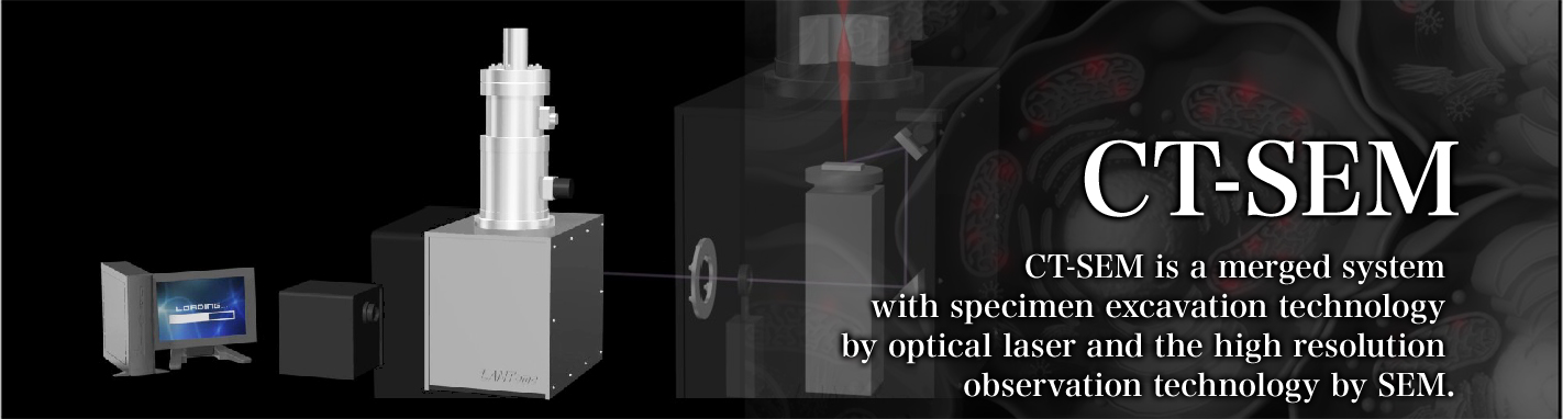
CT-SEM
Features
CT-SEM is the Nano level 3D structural analysis equipment that can reconstruct the acquired SEM image in 3D, while ablating the organic matter surface by the introduction of laser into the sample chamber, with the SEM as a basic body having the resolution of nanometer order. Tomography is conducted by alternately obtaining the surface image of SEM and the ablation of sample surface by laser.
About ablation light source
The sample is not damaged because the greatest feature of this equipment is that it generates very little heat during ablation. The maximum ablation area at one time is 2mm x 2mm, and is very wide-range, and the amount of ablation at one time can be continuously controlled by about 10nm to 400nm by controlling the laser strength at that time. The laser is 100Hz pulse irradiation, and hence the high-speed ablation at a maximum of 40µm per second is enabled.
Ar-F excimer laser is used for the laser source. The wavelength of Ar-F excimer laser is 193nm (6.30eV), and the energy is almost equal to the dissociation energy of C=C double bond. Laser ablation of specimen surface cut the C=C bonds chemically.
- 1. Laser irradiation of energy is almost equal to the C=C bond, which is the main bond in organic objects.
- 2. Excited molecules get photodissociation in pico second order time
- 3. Thermal denaturation is almost free because the ablation energy is consumed for the photodissociation on the specimen surface.
Ablation: the operation such as excluding, cutting, evaporating, elimination, and melting by laser irradiation.
- 1. Dig out the large area rapidly.
- 2. Little thermal denaturation by excavation
- 3. Avoid electron charge-up on insulator surface, because of charge neutralization by electron beam.
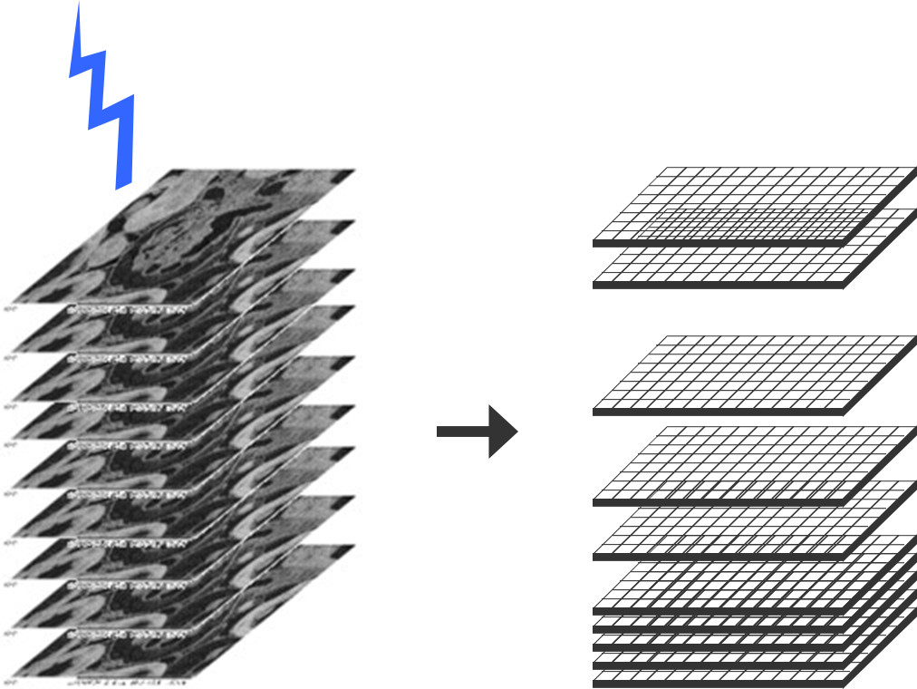
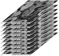
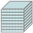
3D data generation flow
For preprocessing, include the biomaterial or softmaterial into epoxy resin, then set them at equipment. Excavation by excimer laser irradiation and imaging of material outer surface by SEM are alternately combined, and SEM images are stored into computer, finally 3D CT model is reconstructed and saved.
Please watch the video for the operation mechanism of CT-SEM